

Click here for the Virus Student Learning Guide
1. Introduction
 Viruses are infectious particles that stand on the border between life and non-life. They’re much smaller and simpler than cells. But, like any living being, they have genes, and that gives them one of the key properties of life: genetic instructions for self-replication. At the same time, viruses lack many of life’s other key properties: they’re not made of cells, and they lack any independent metabolism. Viruses are best described as obligate intracellular parasites, a phrase worth taking apart:
- Parasites are organisms that interact closely with a larger host organism, causing their host harm. While viruses don’t qualify as organisms (an organism is an independent living thing), they certainly prey on a larger host, causing it harm. That host, in some cases, is us. Every time you’ve had a cold, the flu, or another viral infection, you’ve been parasitized by a virus. The COVID-19 pandemic (as we discuss in the next tutorial) was caused by a virus.
- Obligate means “by necessity.” Viruses can’t live on their own. The only way they reproduce themselves is by parasitizing a living cell.
- Intracellular means “within cells.” Viruses are the smallest parasites imaginable, preying upon life at its most basic level: the cell.
While COVID-19 has alerted most people to the importance of viruses, most people aren’t aware of how ubiquitous viruses are. It’s thought that each of Earth’s 9 million or so species is preyed upon by one or more viruses National Library of Medicine. Humans alone might host over 1000 viruses (over 200 have been identified so far). A study at UC Santa Barbara estimated that every liter of seawater might have ten million bacteriophages (viruses that attack bacteria). National Geographic has reported that there might be 1031 viruses on planet Earth: many more than stars in the universe.
So let’s see how these obligate intracellular parasites work. If it suits your learning style, you can start by viewing my music video, I’m a Virus! Otherwise, just continue with the tutorial below.
2. Viruses: Size
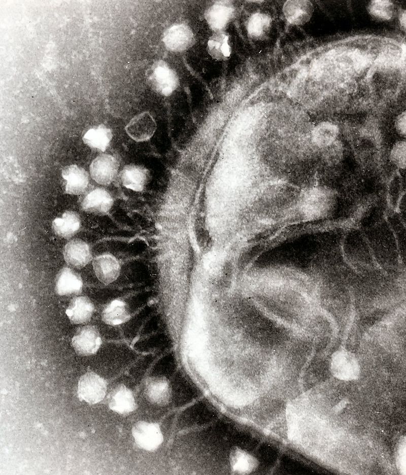

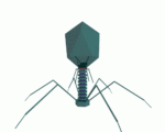
One of the first things to know about viruses is how small they are. The image to the left shows a type of virus called a bacteriophage (or just phage) attached to a bacterial cell. On the right, you can see a comparison between a single bacteriophage and a bacterial cell. Note that the numbers are right, but that visually, the virus should be even smaller. Just to connect the smallness of viruses with what you might already know, think of it this way. A DNA molecule is about 2 nanometers wide. A virus is only about 15 times bigger than that. When we talk about viruses, we’re practically at a molecular scale
3. Viruses: Structure
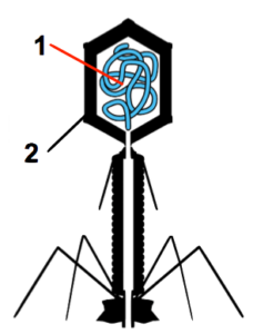
Viruses are also much simpler than cells. To the left, you can see an artist’s depiction of the bacterial viruses that are shown above. These bacterial viruses consist of two main parts: a genetic core (1), consisting of DNA or RNA; and a protein coat (2), also known as a capsid. The capsid, in turn, is made of self-assembling units called capsomeres.
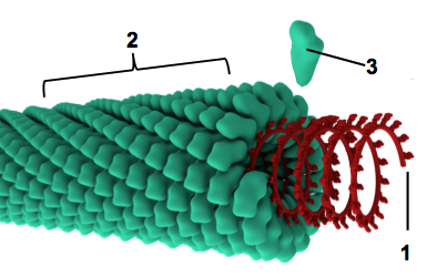
On the right, you can see another type of virus, with the same numbering scheme used to indicate the genetic core (1) and the capsid/protein coat (2). An individual capsomere subunit is at “3.” Note that if the capsomere/capsid relationship is making you think about the tertiary and quaternary levels of structure in proteins, you’ve learned a lot about biology. Good job!
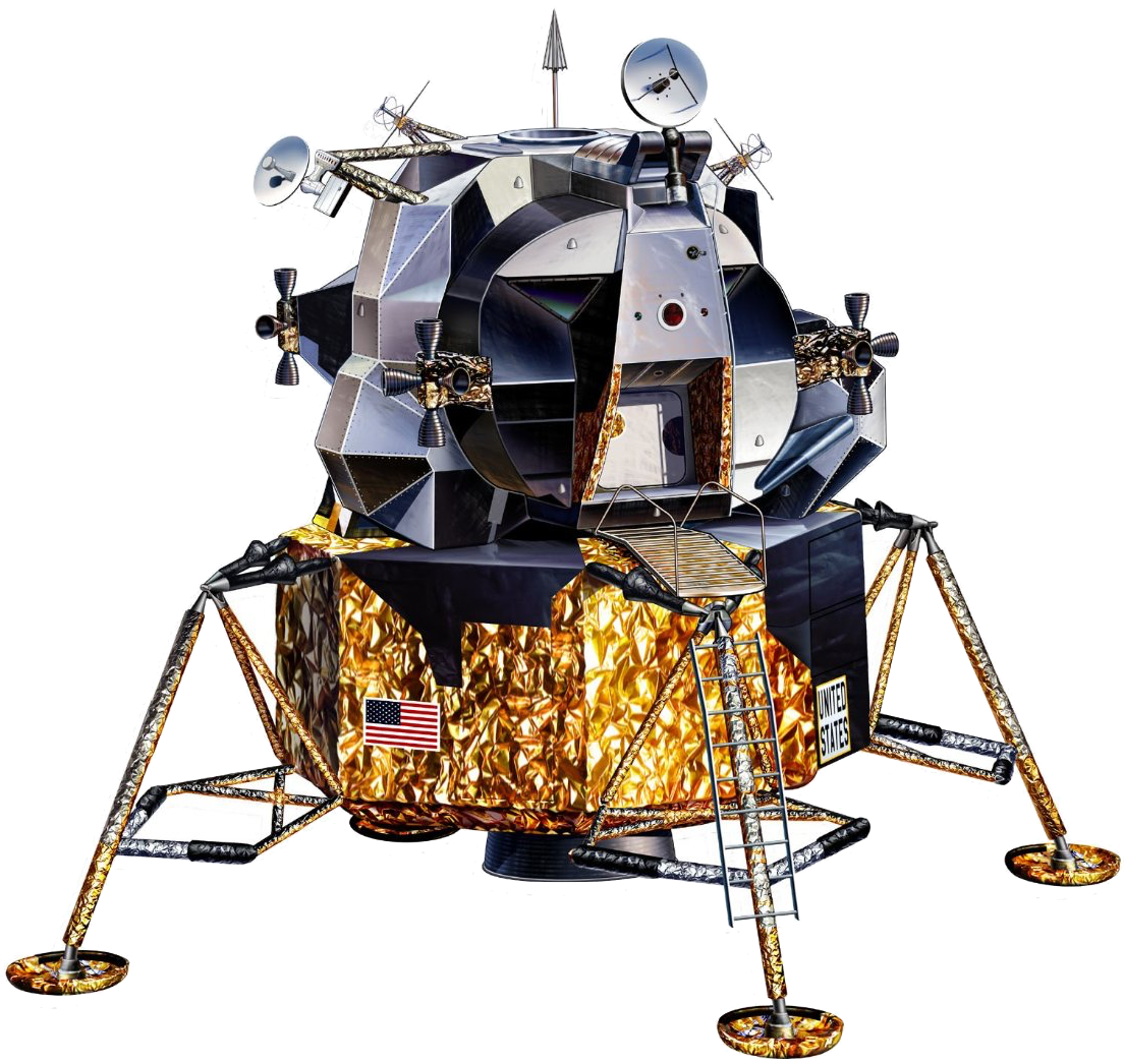
In addition to the capsid, there can also be other protein parts. I’ve always thought that bacteriophages look amazingly similar to NASA’s lunar module, which enabled humans to land on the moon during the Apollo program in the 1960s and 70s. As you can see in the diagram of the bacteriophage above and on the left, this bacteriophage has, on its bottom, what looks like landing gear, and that’s exactly what it is: proteins for attaching to the cells that these viruses parasitize.
Along with landing gear, viruses have various mechanisms for injecting their genes through a cell’s membrane and/or wall, as well as a few other enzymes that the virus will make use of as it attacks its victims.
4. The Lytic Cycle
While viruses aren’t alive, their replication cycle is often referred to as a life cycle. The simplest viral life cycle is called the lytic cycle. In your studies of biology, you’ve met the root lyse several times in words like hydrolysis and lysosome. Hydrolysis is a process in which molecules are broken apart. Lysosomes are organelles that enable cells to digest engulfed food particles (or even organisms). What do you think the lytic cycle breaks apart?
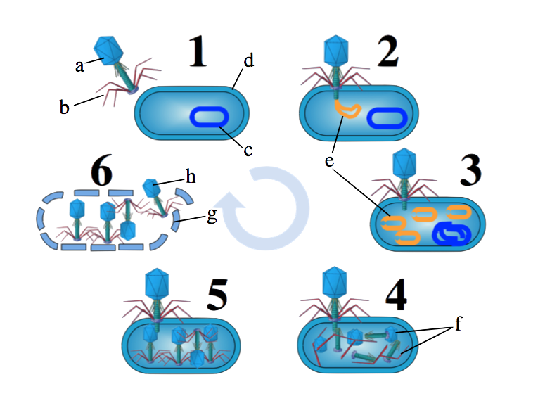 Here’s the lytic cycle for a bacteriophage(or “phage”). As discussed above, that’s the name for a virus that attacks a bacterial cell.
- The cycle begins with a phage (“a”) randomly bumping into its host. Proteins on the phage (the “landing gear” at “b”) enable it to attach to the wall (“d”) of its bacterial host. The host cell’s chromosome is shown at “c.”
- The phage injects its DNA (“e”) into its victim.
- The phage then uses the host cell’s molecular machinery (such as DNA polymerases) to copy the phage’s DNA, while simultaneously shutting down the host cell’s DNA.
- The host’s RNA polymerases and ribosomes are used to create new phage components.
- Based on molecular complementarity, these phage components assemble themselves into new viruses.
- At a certain point, the host is so stuffed with phages that it bursts, releasing new phages into the environment.
Got it? Label the diagrams below.
[qwiz qrecord_id=”sciencemusicvideosMeister1961-Viruses and the Lytic Cycle (M15)”]
[h]Viruses and the Lytic Cycle (labeled diagram quiz)
[i]
[q labels = “right”]
[l]genes (DNA or RNA)
[fx] No. Please try again.
[f*] Correct!
[l]protein
[fx] No. Please try again.
[f*] Excellent!
[q labels = “top”]
[l]genes
[fx] No. Please try again.
[f*] Correct!
[l]capsid
[fx] No, that’s not correct. Please try again.
[f*] Correct!
[l]capsomere
[fx] No. Please try again.
[f*] Great!
[q labels = “top”]
[l]attachment
[fx] No. Please try again.
[f*] Excellent!
[l]injection of viral DNA
[fx] No, that’s not correct. Please try again.
[f*] Good!
[l]lysis
[fx] No. Please try again.
[f*] Good!
[l]replication of viral DNA
[fx] No. Please try again.
[f*] Excellent!
[l]self-assembly of new viruses
[fx] No, that’s not correct. Please try again.
[f*] Excellent!
[l]synthesis of viral proteins
[fx] No. Please try again.
[f*] Correct!
[/qwiz]
5. The Lysogenic Cycle
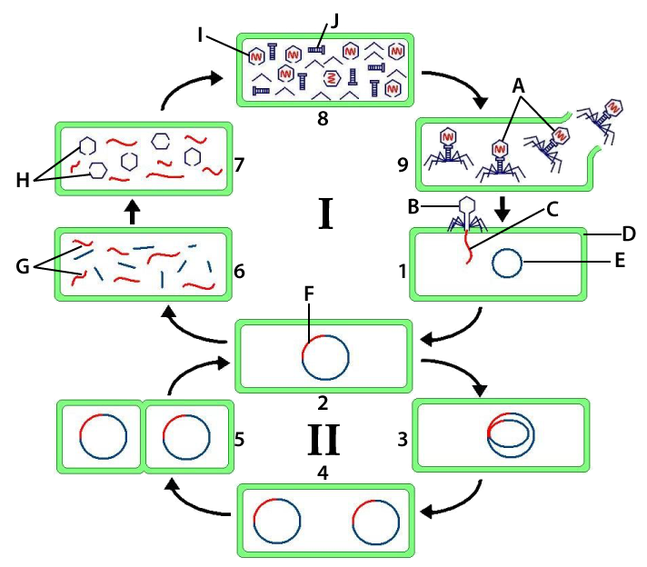
In the lysogenic cycle, viral genes incorporate themselves into the chromosomes of their hosts. As you can see in image 1 (on the upper right side of the diagram to your left), the cycle begins just as the lytic cycle does: with the virus (B) injecting its genes (C) into its host (D).
But in step two, things start to differ. Here, the viral DNA (in red) incorporates itself into its host’s chromosome (at “E,” in blue). Once there, the viral DNA is called a provirus (or a prophage, if it’s a bacterial virus).
From this point on, as you can see in part II of the diagram (images 3, 4, and 5) the provirus becomes part of (and benefits from) the host cell’s reproductive cycle. In other words, every time the cell replicates itself, the cell will also replicate the provirus. Since bacteria can reproduce themselves very quickly (E. coli can reproduce itself every 20 minutes under ideal conditions), lysogenic incorporation can be an effective reproductive strategy for lysogenic viruses.
At certain moments, however, proviruses can break out of their dormancy, excise themselves from their host chromosome, and re-enter the virulent, lytic cycle. It’s possible that certain environmental stimuli, such as exposure to ultraviolet light, induce this change Encyclopedia Britannica. The consequences of this break-out are seen in the top half of the diagram: in # 6, viral genes are synthesized. In # 7, viral proteins are translated. In # 8, self-assembly of viral parts occurs, leading to lysis in step 9.
Use what you’ve learned above, plus your knowledge of biology, to label the diagram below.
[qwiz qrecord_id=”sciencemusicvideosMeister1961-Lysogenic Viruses Interactive Diagram”]
[h]The Lysogenic Cycle: Interactive Diagram
[q labels = “top”]
[l]binary fission
[fx] No, that’s not correct. Please try again.
[f*] Good!
[l]provirus break out: production of viral DNA
[fx] No. Please try again.
[f*] Correct!
[l]DNA replication, including phage DNA
[fx] No. Please try again.
[f*] Excellent!
[l]injection of viral genes
[fx] No. Please try again.
[f*] Excellent!
[l]incorporation of phage DNA into host chromosome
[fx] No. Please try again.
[f*] Good!
[l]lysis
[fx] No. Please try again.
[f*] Correct!
[l]production of viral proteins
[fx] No. Please try again.
[f*] Great!
[l]assembly of new viral parts
[fx] No. Please try again.
[f*] Excellent!
[x][restart]
[/qwiz]
6. HIV: Structure and Life Cycle
“HIV” is an acronym for Human Immunodeficiency Virus. Infection with HIV, if untreated, causes AIDS: acquired immunodeficiency syndrome. Let’s look at that term:
- Acquired: you have to be infected with the virus to get AIDS. In other words, unlike some other disorders of the immune system, it’s not inherited.
- Immunodeficiency. Your immune system is how your body fights off invading disease-causing organisms such as bacteria or fungi, as well as viruses. If you’re immunodeficient, you lack all or some of your body’s capacity to fight off these invaders.
Here’s a description of how HIV causes AIDS from HIV.gov
HIV attacks the body’s immune system, specifically the CD4 cells (T cells), which help the immune system fight off infections. If left untreated, HIV reduces the number of CD4 cells (T cells) in the body, making the person more likely to get infections or infection-related cancers. Over time, HIV can destroy so many of these cells that the body can’t fight off infections and disease. These opportunistic infections or cancers take advantage of a very weak immune system and signal that the person has AIDS, the last state of HIV infection.
In terms of our AP Biology course, one of the key things to understand about HIV is that it’s a retrovirus. Retroviruses have RNA genes, and then use an enzyme called reverse transcriptase to make a DNA transcript of their RNA. Note that this is the opposite of what usually happens during transcription, during which DNA is transcribed into RNA: that’s why this is reverse transcription, and why HIV is a retrovirus. This DNA then lysogenizes, entering into the nuclear DNA of its host, the helper T cell.
Let’s start by looking at the key parts of HIV’s structure, and then we’ll go through its life cycle.
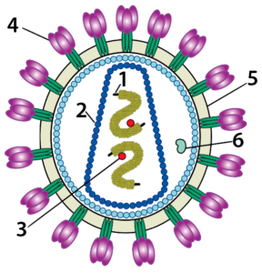
“1” is HIV’s RNA genome. Surrounding the genes (at “2”) is a capsid. “3” is reverse transcriptase, an enzyme whose function is described immediately above. “4” indicates the glycoproteins (proteins with an attached carbohydrate) that enable HIV to bind with and inject its genes and enzymes into its host. These glycoproteins are embedded in a phospholipid bilayer (“5”). As we’ll see below, these phospholipids are stolen from the infected helper T cell as HIV oozes out of its victim in a process that closely resembles exocytosis. “6” is another enzyme called “protease.” Protease modifies some of the viral polypeptides that HIV forces its host to synthesize, creating mature viral proteins that can be assembled into new viruses.
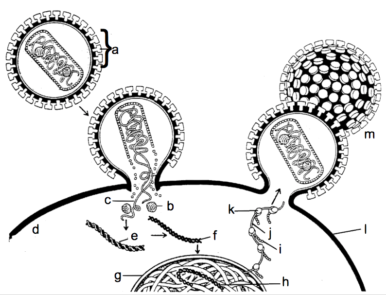
Let’s now look at HIV’s life cycle. The letter “a” represents HIV in an infected person’s bloodstream. The glycoproteins on HIV’s surface enable it to bind with membrane proteins on the surface of HIV’s target, the Helper T cell (“d”). Binding leads to the fusion of HIV with the Helper T, allowing viral RNA (“c”) and reverse transcriptase (“b”) to enter the cell.
The function of reverse transcriptase is to convert the information in viral RNA into DNA. Using HIV’s RNA as a template, reverse transcriptase creates a hybrid DNA/RNA molecule (shown at “e”), which is then converted to a molecule that is purely viral DNA (at “f”). This viral DNA then enters the Helper T cell’s nucleus (“g”), where it becomes incorporated into the T cell’s chromosomal DNA as a provirus (“H”). Once in the nucleus, HIV’s DNA will be transcribed into RNA (“j”). The cell’s ribosomes (“k”) will translate the viral RNA into viral proteins (“i”), which will self-assemble into new viruses (“m”) that bud off from the helper T cell’s membrane (“l”), to be released into the blood or lymph.
Take this quiz to make sure you’re understanding how HIV works.
[qwiz qrecord_id=”sciencemusicvideosMeister1961-HIV Interactive Diagrams (M15) ” style=”width: 600px !important; min-height: 400px !important;”]
[h] HIV: Interactive Diagrams
[i]
[q labels = “top”]
[l]capsid
[f*] Great!
[fx] No, that’s not correct. Please try again.
[l]glycoprotein
[f*] Excellent!
[fx] No. Please try again.
[l]phospholipid bilayer
[f*] Good!
[fx] No. Please try again.
[l]protease
[f*] Good!
[fx] No. Please try again.
[l]reverse transcriptase
[f*] Excellent!
[fx] No, that’s not correct. Please try again.
[l]RNA
[f*] Correct!
[fx] No. Please try again.
[q labels = “top”]
[l]docks with Helper T cell
[f*] Good!
[fx] No. Please try again.
[l]genes
[f*] Great!
[fx] No. Please try again.
[l]makes DNA from RNA
[f*] Good!
[fx] No. Please try again.
[l]modifies HIV proteins
[f*] Good!
[fx] No. Please try again.
[l]protein coat
[f*] Excellent!
[fx] No, that’s not correct. Please try again.
[l]stolen from host cell membrane
[f*] Excellent!
[fx] No. Please try again.
[q labels = “top”]
[l]cytoplasm of Helper T
[f*] Excellent!
[fx] No. Please try again.
[l]Helper T membrane
[f*] Correct!
[fx] No, that’s not correct. Please try again.
[l]HIV Glycoprotein
[f*] Good!
[fx] No. Please try again.
[l]hybrid viral DNA-RNA
[f*] Correct!
[fx] No, that’s not correct. Please try again.
[l]nucleus
[f*] Good!
[fx] No, that’s not correct. Please try again.
[l]provirus
[f*] Good!
[fx] No, that’s not correct. Please try again.
[l]reverse transcriptase
[f*] Correct!
[fx] No. Please try again.
[l]ribosome
[f*] Correct!
[fx] No, that’s not correct. Please try again.
[l]viral RNA
[f*] Correct!
[fx] No. Please try again.
[l]viral protein
[f*] Excellent!
[fx] No. Please try again.
[l]new virus
[f*] Correct!
[fx] No, that’s not correct. Please try again.
[l]viral DNA
[f*] Correct!
[fx] No, that’s not correct. Please try again.
[/qwiz]
7. Viruses and Variation
Along two different axes, viruses can be thought of as engines of variation. On the one hand, viruses generate diversity within their own populations. This diversity arises from
- Mutations that occur during viral replication. The rate of mutation in DNA viruses is similar to that of eukaryotic cells. However, the mutation rate in RNA viruses can be significantly higher. Medical Microbiology
- Mixing of viral genomes. When two viruses invade the same host cell, the resulting viral offspring can wind up with recombined DNA from the two invading viruses. This type of recombination in the influenza virus, which can infect humans, birds, and pigs, explains the virus’s variability from year to year (and the consequent need for the development of a new influenza vaccine each year). Medical Microbiology
In addition, viruses generate diversity within their hosts. This process is known as transduction. There are two forms of transduction: specialized and generalized. For AP biology, understanding generalized transduction is sufficient. If you want to learn about specialized transduction, click here to read a Wikipedia article about it).
![]() In generalized transduction, mistakes during the lytic cycle result in the transfer of genes between bacterial cells. You can see how this works in the series of diagrams to your left. In Diagram # 1, a phage (a) attacks a bacterial cell, injecting its viral DNA (b). The host cell’s chromosome is indicated by the letter “c.” Diagram 2 shows the lytic cycle progressing, with the cell making copies of the virus’s DNA (“b”) as its own chromosome disintegrates into pieces of DNA (“d”).
In generalized transduction, mistakes during the lytic cycle result in the transfer of genes between bacterial cells. You can see how this works in the series of diagrams to your left. In Diagram # 1, a phage (a) attacks a bacterial cell, injecting its viral DNA (b). The host cell’s chromosome is indicated by the letter “c.” Diagram 2 shows the lytic cycle progressing, with the cell making copies of the virus’s DNA (“b”) as its own chromosome disintegrates into pieces of DNA (“d”).
Diagram # 3 shows the mistake that’s at the heart of the process of generalized transduction. Three phages are shown in this diagram. The phage indicated by the letter “e” is carrying viral DNA (indicated by the red-colored DNA strand inside its capsid). By contrast, the phage indicated by the letter “f” picked up the wrong DNA. Instead of viral DNA, this phage incorporated a strand of DNA from the first viral host (host # 1).
Diagram # 4 shows what will happen if phage “f” attacks another host (host # 2). Instead of injecting viral DNA, phage “f” will inject the DNA from host # 1.
In diagram # 5, we see the result. Various enzymes called recombinases will line up the new DNA (from host # 1) with the DNA from host # 2, and recombine the two, swapping segments of DNA between them. In this case, the result is a bacterial chromosome that combines DNA from host # 1 with host # 2. In other words, transduction has created a chromosome with a novel DNA sequence.
8. I’m a Virus: Interactive Lyrics
Here’s much of what you need to know about viruses, in a matching quiz format with rhyming lyrics! Holy Cow!
[qwiz qrecord_id=”sciencemusicvideosMeister1961-I’m a Virus, Interactive Lyrics”]
[h]Virus Interactive Lyrics
[i]
[q labels = “top”]
I’m a virus, an _________ particle,
a nano-thug, a pirate, the genuine article
Ebola, chickenpox, west Nile, influenza,
Yellow fever, AIDS, herpes, SARS, I’m comin’ right at ya
I’m not a _____, not an independent organism,
I don’t even have my own __________,
I only __________ myself by taking over cells,
Then I bust ‘em apart, no wonder you don’t feel well!
[l]cell
[fx] No. Please try again.
[f*] Correct!
[l]infectious
[fx] No. Please try again.
[f*] Excellent!
[l]metabolism
[fx] No, that’s not correct. Please try again.
[f*] Excellent!
[l]reproduce
[fx] No, that’s not correct. Please try again.
[f*] Good!
[q labels = “top”]
My structure is simple, mostly ________ and genes,
Packaged as a sub-micron killing machine,
My ________ made of capsomeres that self _________,
Whenever cells see me they start to tremble,
Inside my capsid is where my ______ reside,
I got DNA or _____ on the inside,
And the rest of me is various proteins whose role,
Is assisting in assaulting cells and taking control.
CHORUS
I’m a virus,
Gonna use your cells to ________ me
I’m a virus
The death of your cells is the life of me
[l]assemble
[fx] No. Please try again.
[f*] Excellent!
[l]capsid’s
[fx] No, that’s not correct. Please try again.
[f*] Excellent!
[l]genes
[fx] No, that’s not correct. Please try again.
[f*] Good!
[l]proteins
[fx] No, that’s not correct. Please try again.
[f*] Excellent!
[l]replicate
[fx] No. Please try again.
[f*] Excellent!
[l]RNA
[fx] No, that’s not correct. Please try again.
[f*] Great!
[q labels = “top”]
The _____ cycle shows one method of attack,
I attach to the cell surface and pierce it like a tack
Inject my ______ inside, use my victim’s __________,
to make my genes and proteins for reproducing more of me.
Step 1, __________ grab the membrane or the wall
2: inject my genes, this cell is gonna fall.
3: use the victim’s __________ and ribosomes,
To synthesize my proteins and replicate my genome.
4: tailpiece and capsid self-assembly,
5: I’m all together now, look at all my progeny
6 is lysis, I burst that _____ apart,
Now I’m looking for more victims I can dearly depart.
CHORUS
[l]cell
[fx] No, that’s not correct. Please try again.
[f*] Excellent!
[l]genes
[fx] No. Please try again.
[f*] Great!
[l]lytic
[fx] No, that’s not correct. Please try again.
[f*] Excellent!
[l]machinery
[fx] No, that’s not correct. Please try again.
[f*] Great!
[l]polymerase
[fx] No, that’s not correct. Please try again.
[f*] Great!
[l]tail fibers
[fx] No, that’s not correct. Please try again.
[f*] Great!
[q labels = “top”]
Sometimes instead of lysing I __________
Which happens after I inject my genes inside
I slip into your ___________ a thief inside your room
A menacing and silent presence bringer of doom
Integrated in you, your ______ my home,
And see what happens when you replicate your chromosomes
Each copy that you copy copies me as well as you,
You might make a million ______, there’s nothin’ you can do
[l]chromosome
[fx] No, that’s not correct. Please try again.
[f*] Excellent!
[l]copies
[fx] No. Please try again.
[f*] Correct!
[l]DNA’s
[fx] No, that’s not correct. Please try again.
[f*] Good!
[l]lysogenize
[fx] No. Please try again.
[f*] Correct!
[q labels = “top”]
My lurking hiding being’s called a _________ or provirus,
And if you got another name just call us or wire us,
Or write it on papyrus ‘til the moment that I emerge,
from your DNA I’m _____ now a cell-destroying scourge.
And suddenly your cells are _____ watch me as I lyse them,
I’m a pirate so forget your plans my program now denies them,
Your cells are _____ factories they’re making millions more of me
I leave your cells and now I’m free Infecting you’s my destiny
CHORUS
[l]dying
[fx] No. Please try again.
[f*] Good!
[l]lytic
[fx] No, that’s not correct. Please try again.
[f*] Correct!
[l]prophage
[fx] No. Please try again.
[f*] Excellent!
[l]virus
[fx] No. Please try again.
[f*] Great!
[q labels = “top”]
Now meet my associate the virus ______
Who’s helpless target is the cell called “T”
HIV causes ________________,
Raising cancer and infection vulnerability
Remember that the T’s an essential cell
In the _______ system army it’s the general
So knocking out the ______ sets the stage
For cancers, germs, and viruses like me to invade!
[l]HIV
[fx] No. Please try again.
[f*] Great!
[l]immune
[fx] No, that’s not correct. Please try again.
[f*] Excellent!
[l]immunodeficiency
[fx] No. Please try again.
[f*] Great!
[l]T-cell
[fx] No. Please try again.
[f*] Good!
[q labels = “top”]
HIV’s genes are made of _____,
Outside its capsid’s a ___________ membrane,
A skin stolen from the cell it last dispatched,
studded with proteins that let it attach,
To a protein on the _____ called CD4
Inducing that cell to open up its door,
Inviting in _____ enzymes and core,
That T-cells already done for.
[l]HIV’s
[fx] No, that’s not correct. Please try again.
[f*] Good!
[l]phospholipid
[fx] No, that’s not correct. Please try again.
[f*] Correct!
[l]RNA
[fx] No, that’s not correct. Please try again.
[f*] Correct!
[l]T-cell
[fx] No. Please try again.
[f*] Excellent!
[q labels = “top”]
HIV’s a __________ it’s next routine,
has to do with transforming its RNA genes
___________________ takes its RNA
And reverse transcribes it into _____
Then this DNA integrates into your ______________,
Like a __________ prophage in your genome.
This proviral DNA’s __________ and translated
As new virus genes and proteins get created
And like any virus, all those parts will self _________,
Into new viruses which of course resemble,
the original particle which started the infection,
Acquired by sex, or a needle injection
[l]assemble
[fx] No, that’s not correct. Please try again.
[f*] Excellent!
[l]chromosomes
[fx] No, that’s not correct. Please try again.
[f*] Correct!
[l]DNA
[fx] No, that’s not correct. Please try again.
[f*] Correct!
[l]lysogenic
[fx] No, that’s not correct. Please try again.
[f*] Good!
[l]retrovirus
[fx] No, that’s not correct. Please try again.
[f*] Excellent!
[l]reverse transcriptase
[fx] No, that’s not correct. Please try again.
[f*] Correct!
[l]transcribed
[fx] No, that’s not correct. Please try again.
[f*] Great!
[/qwiz]
9. A virus quiz
If you understand 1) viral structure, 2) the lytic cycle, 3) the lysogenic cycle, 4) the life cycle of retroviruses like HIV, and 5) how viruses act to increase variation both within themselves and their hosts, then you know everything about this topic that’s required for an AP Biology course. To test your understanding, take the quiz below (which mostly focuses on the diagrams above).
[qwiz random = “true” dataset_intro=”false” use_dataset=”Viruses Dataset” qrecord_id=”sciencemusicvideosMeister1961-Virus Cumulative Quiz”]
[h]Virus Cumulative Quiz
[i]
[x][restart]
[/qwiz]
10. Next Steps
Continue on to Understanding SARS-CoV-2, the next tutorial in this module.